The goal of ravetools is to provide memory-efficient signal & image processing toolbox for intracranial Electroencephalography. Highlighted features include:
-
Notch filter(remove electrical line frequencies) -
Welch Periodogram(averaged power over frequencies) -
Wavelet(frequency-time decomposition) - 2D, 3D image convolution via
FFT -
CT/MRItoMRIimage alignment
Installation
The package is available on CRAN. To install the compiled version, simply run:
install.packages("ravetools")Installing the package from source requires installation of proper compilers and some C libraries; see this document for details.
iEEG preprocess pipeline
This is a basic example which shows you how to preprocess an iEEG signal. The goal here is to:
- Plot diagnostic graphs to inspect channels
- Apply Notch filters to remove electrical line noise
- Frequency-time decomposition and show the power densities
* Channel referencing is not included
1. Generate toy examples:
library(ravetools)

# Generate 20 second data at 2000 Hz
time <- seq(0, 20, by = 1 / 2000)
signal <- sin( 120 * pi * time) +
  sin(time * 20*pi) +
  exp(-time^2) *
  cos(time * 10*pi) +
  rnorm(length(time))

diagnose_channel(signal, srate = 2000)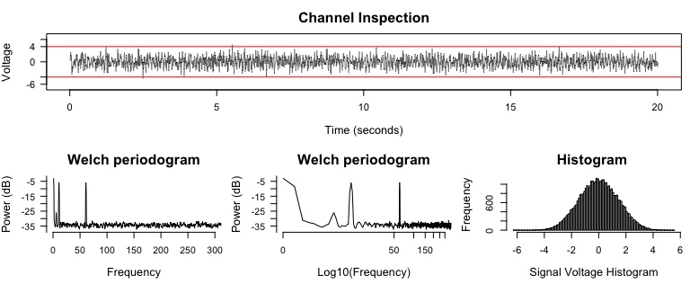
2. Apply Notch filters and inspect Periodograms
## ------- Notch filter --------
signal2 <- notch_filter(signal, sample_rate = 2000)

diagnose_channel(signal, signal2, srate = 2000,
                 name = c("Raw", "Filtered"))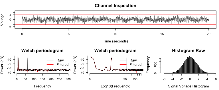
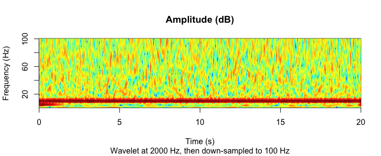
3. Frequency-time decomposition
Current version of ravetools provides two approaches: Wavelet and Multi-taper. Wavelet uses the Morlet wavelet and obtains both amplitude and phase data, while Multi-taper does not generate phase data. However, the amplitude obtained from Multi-taper is smoother than Wavelet.
Using Wavelet:
## ---------- Wavelet -----------
coef <- morlet_wavelet(
  signal2, freqs = seq(1, 100, by = 1),
  srate = 2000, wave_num = c(2, 15))
amplitude <- 20 * log10(Mod(coef[]))

# For each frequency, decimate to 100 Hz
downsample_amp <- apply(amplitude, 2, decimate, q = 20)
downsample_time <- decimate(time, q = 20)

par(mfrow = c(1,1))
image(
  z = downsample_amp,
  x = downsample_time,
  y = seq(1, 100, by = 1),
  xlab = "Time (s)",
  ylab = "Frequency (Hz)",
  main = "Amplitude (dB)",
  sub = "Wavelet at 2000 Hz, then down-sampled to 100 Hz",
  col = matlab_palette()
)
Multi-taper
Alternatively you can use Multi-tapers to obtain amplitude data. The algorithm is modified from source code here. Please credit them as well if you adopt this approach.
## ---------- Multitaper -----------
res <- multitaper(
  data = signal2,
  fs = 2000,
  frequency_range = c(1, 100),
  time_bandwidth = 1.5,
  window_params = c(2, 0.01),
  nfft = 100
)

par(mfrow = c(1,1))
image(
  x = res$time,
  y = res$frequency,
  z = 10 * log10(res$spec),
  xlab = "Time (s)",
  ylab = 'Frequency (Hz)',
  col = matlab_palette(),
  main = "Amplitude (dB)"
)
Image alignment
ravetools provides imaging co-registration via NiftyReg (doi.org/10.1117/1.JMI.1.2.024003). You can align CT to MRI, or MRI (T2) to MRI (T1). The method can be body rigid, affine, or non-linear.
source <- system.file("extdata", "epi_t2.nii.gz", package="RNiftyReg")
target <- system.file("extdata", "flash_t1.nii.gz", package="RNiftyReg")
aligned <- register_volume(source, target, verbose = FALSE)

source_img <- aligned$source[[1]]
target_img <- aligned$target
aligned_img <- aligned$image

par(mfrow = c(2, 2), mar = c(0.1, 0.1, 3.1, 0.1))

pal <- grDevices::grey.colors(256, alpha = 1)
image(source_img[,,30], asp = 1, axes = FALSE,
      col = pal, main = "Source image")
image(target_img[,,64], asp = 1, axes = FALSE,
      col = pal, main = "Target image")
image(aligned_img[,,64], asp = 1, axes = FALSE,
      col = pal, main = "Aligned image")

# bucket fill and calculate differences
aligned_img[is.nan(aligned_img) | aligned_img <= 1] <- 1
target_img[is.nan(target_img) | aligned_img <= 1] <- 1
diff <- abs(aligned_img / target_img - 1)
image(diff[,,64], asp = 1, axes = FALSE,
      col = pal, main = "Percentage Difference")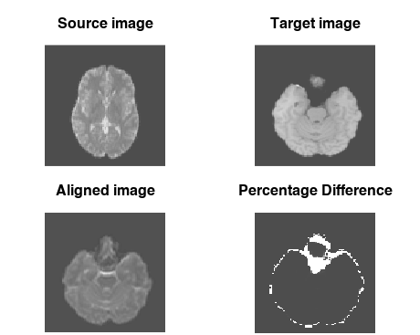
References
To cite ravetools in publications use, please cite the RAVE paper from Beauchamp's lab
Magnotti, JF, and Wang, Z, and Beauchamp, MS. RAVE: comprehensive
  open-source software for reproducible analysis and visualization of
  intracranial EEG data. NeuroImage, 223, p.117341.
The multitaper function (MIT License) uses the script derived from Prerau's lab. The TinyParallel script is derived from RcppParallel package (GPL License) with TBB features removed (only use tinythreads). The register_volume function uses NiftyReg (BSD License) developed by CMIC at University College London, UK (its R implementation is released under GPL license).
[1] Magnotti, JF, and Wang, Z, and Beauchamp, MS. RAVE: comprehensive
    open-source software for reproducible analysis and visualization of
    intracranial EEG data. NeuroImage, 223, p.117341.
[2] Prerau, Michael J, and Brown, Ritchie E, and Bianchi, Matt T, and
    Ellenbogen, Jeffrey M, and Purdon, Patrick L. Sleep Neurophysiological
    Dynamics Through the Lens of Multitaper Spectral Analysis. Physiology,
    December 7, 2016, 60-92.
[3] Modat, M., Cash, D.M., Daga, P., Winston, G.P., Duncan, J.S. and
    Ourselin, S., 2014. Global image registration using a symmetric
    block-matching approach. Journal of medical imaging, 1(2), pp.024003-024003.
[4] JJ Allaire, Romain Francois, Kevin Ushey, Gregory Vandenbrouck, Marcus
    Geelnard and Intel (2022). RcppParallel: Parallel Programming Tools for
    'Rcpp'. R package version 5.1.5.
    https://CRAN.R-project.org/package=RcppParallel